Root canal treatment is often used to save a tooth that has been damaged or is severely decayed. The root canal is situated in the middle of the tooth and contains the living tissue of the tooth, known as the pulp. If the pulp should become infected, the tooth will effectively start to die and, if the tooth is left untreated, the tooth will eventually become rotten and may need to be extracted. Root canal treatment involves taking out the infected pulp and cleaning the tooth, which is then sealed and fitted with a crown or other suitable restorative option.
Root canal treatment can be carried out by general or specialist dentists, but general dentists tend to refer complex cases to specialist dentists who are known as endodontists.
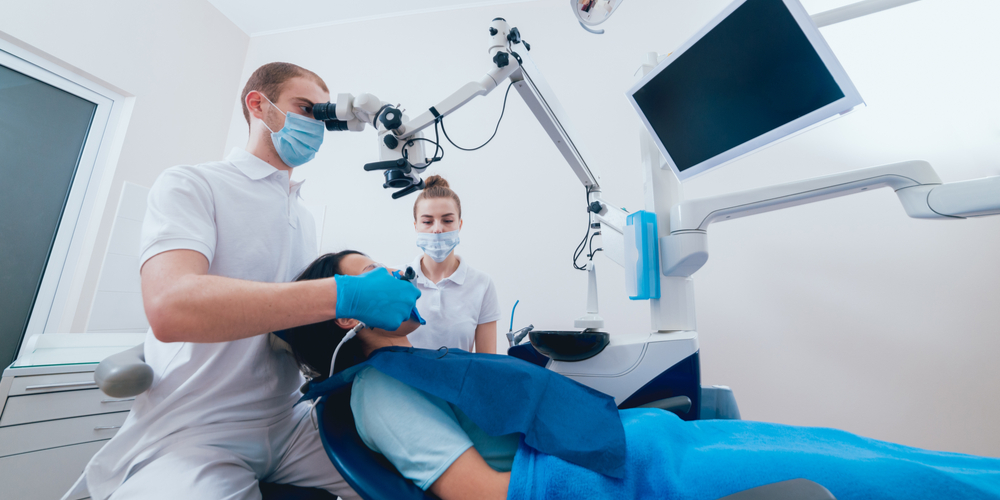
How are dental microscopes used in endodontics?
Research has revealed that endodontists value dental microscopes as a very important tool. A study carried out in 2004 found that 68 percent of endodontists regularly use a dental microscope as part of the treatment process. Dental microscopes have the following benefits for endodontists:
- improved lighting
- makes it easier to detect additional canals
- improves the accuracy of incisions and suturing
- allows the specialist to reduce the size of the surgical site, which can speed up the healing process
- facilitates the cleaning of the canals
- facilitates the detection of abnormalities or root fractures
- provides a better operating position, which helps to eliminate discomfort and back pain
- locating calcified canals
It is vital for dentists to be able to see during endodontic treatment, so the illumination and magnification produced by a microscope can prove invaluable.
The microscope may also be beneficial for patients because dentists can explain the treatment more clearly, which can help the patient feel more comfortable about the procedure.