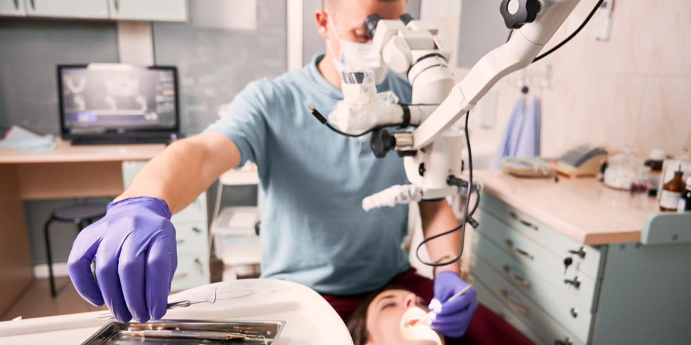
Periodontal dentistry is concerned with the diagnosis and treatment of conditions affecting the gum tissue. It is a serious form of gum disease, which can result in irreversible damage to the teeth and gums if not properly treated. Gum disease is one of the most common preventable illnesses in society, but it is also one that can be completely treated in the majority of cases. However, if the disease should be left to develop, the gums may become very painful and swollen and the teeth may eventually come loose.
In order to prevent gum disease, it is important to adopt a good daily oral hygiene routine, which includes brushing the teeth twice a day, using dental floss and mouthwash, and attending regular check-ups with your dentist, who can identify early signs of gum disease that may not be noticeable to you.
Signs and symptoms of gum disease that you should look out for include sore and swollen gums, bleeding gums, heightened sensitivity and red gums.
How are dental microscopes used in periodontal dentistry?
Dental microscopes have many different functions in aiding the treatment of periodontal problems. The benefits of using a dental microscope in this specialist area are numerous, including:
- allows dentists to reduce the size of the surgical site, which can speed up the healing process
- increases the accuracy of surgical incisions and suturing
- facilitates better inspection and evaluation of soft tissue lesions in the mouth
- facilitates the detection of micro-inflammation
- improves the evaluation of restorations and marginal tissue
- magnifies and illuminates the root surface for greater visibility
- improves control of laser surgery
- enables micro-level osseous surgery
- facilitates sinus-lift operations
- allows dissection of the mental and mandibular nerves
